September 23, 2021 | Net Health
3 Minute Read
Help Your Patients Tell the Best Wound Care Story Possible
Simple tips on taking better photos to share with clinicians
They say a picture is worth a thousand words, and in the world of wound care, that statement is especially true, especially now.
During the pandemic, and now with the uncertainty of Delta and other variants, 2D and 3D images taken on digital devices are helping clinicians stay connected to their patients.
Why Take Images?
Photos of patients’ wounds provide a range of vital information, including:
• Wound size
• Wound depth
• Wound tissue and amount (granulation, slough, eschar, epithelial)
• Quality of wound edges and the periwound area (maceration, epibole, shape)
• Pre- and post-debridement wound status, to validate the effectiveness
• Timeline of healing
There is now a handful of wound care mobile apps that help patients and clinicians capture images and send them back to the wound care specialists for analysis and treatment. One of the leading mobile wound care apps for patients is Tissue Analytics (TA), a Net Health company. TA introduced an imaging app for patients in 2020 to help keep providers and patients engaged during the pandemic.
With the company’s technology, patients get a push alert. They are then prompted to send a snapshot of the wound, and answer a short survey. Nurses or other clinicians receive the data and review it asynchronously when that clinician has time. The data is also be uploaded in the EHR, where it can become an important part of the patient record.
5 Tips for Better Wound Care Images
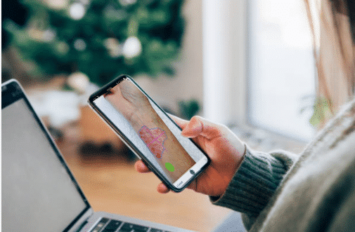
The process of keeping wound care patients electronically engaged with their providers starts with getting a clear, crisp image of the wound. And that process is more than just point and click. Here’s a list of 5 simple tips to help patients using Tissue Analytics app take the best possible pictures.
1. Use a smartphone camera. Most Android and iOS smartphones today provide excellent photographic technology and most patients (or their family members) will have a smartphone.
2. Provide the proper perspective. One of the most critical steps to take when taking an image of a wound is to ensure you have the appropriate perspective, so the size of the wound is clear to see. Our Tissue Analytics platform uses a green adhesive dot placed near the wound. This allows the clinicians to analyze the image to get the true size of the wound area. (Note – the dot should not touch the wound, there should be some healthy tissue between the wound and dot. The dot should not be at an angle – but on the same plane as the wound.)
3. Ensure great lighting. Don’t take pictures in a dark room, and always make sure the flash is on. Note – don’t rely on the automatic flash feature – manually turn on the flash. Likewise, make sure there is no glare if you are in direct sunlight. Take a few experimental shots to be sure!
4. Make sure you aren’t too far away. If you take the image from a distance, the camera won’t get the details the clinician needs. Patients should get as close as possible to show the entire wound area – side to side and up and down.
5. Get the right angle. Hold device parallel to the wound, so the picture looks flat – like you are looking at the wound face on. Avoid taking photos at an angle as the perspective will be disjointed.
Clinicians may remember that learning how to use a mobile app took them a few tries as well. Be patient and help your patients learn best practices so that you and them get the benefits of optimal wound care treatments.
Wound care images help with outcomes and quality of life!

Top 5 Digital Techniques to Reduce Hospital-Acquired Pressure Injuries
Prevent HAPIs and provide better overall care